Tempowire surgical temporary heart pace wire has one-edge slipped insulation or diameter. It is fastened to myocardium with curved, round suture needle connected to ensure conductivity or fastened with a anchor by passing through the myocardium if it has anchor. The anchor is the stripped and winged piece of insulation close to the curved needle. The other end of the insulated wire is connected to the straight, sharp Keith needle that enables passage from inside to outside from the chest cavity wall to ensure conductivity. Keith needle is equipped with a special notch ensuring frangibility. The sharp part of the surgical needle is broken off from the notch, the part that remains attached to the wire forms the lead pin and connects to conventional pace or monitor equipment.
Usage Techniques for Temporary Pace Wire
Knowledge Base
What are the things you need to know about the low density PE (LD-PE) coated (insulated) Tempowire surgical temporary heart pace wire and what are the usage techniques?
Tempowire Surgical Temporary Pace Wire Usage

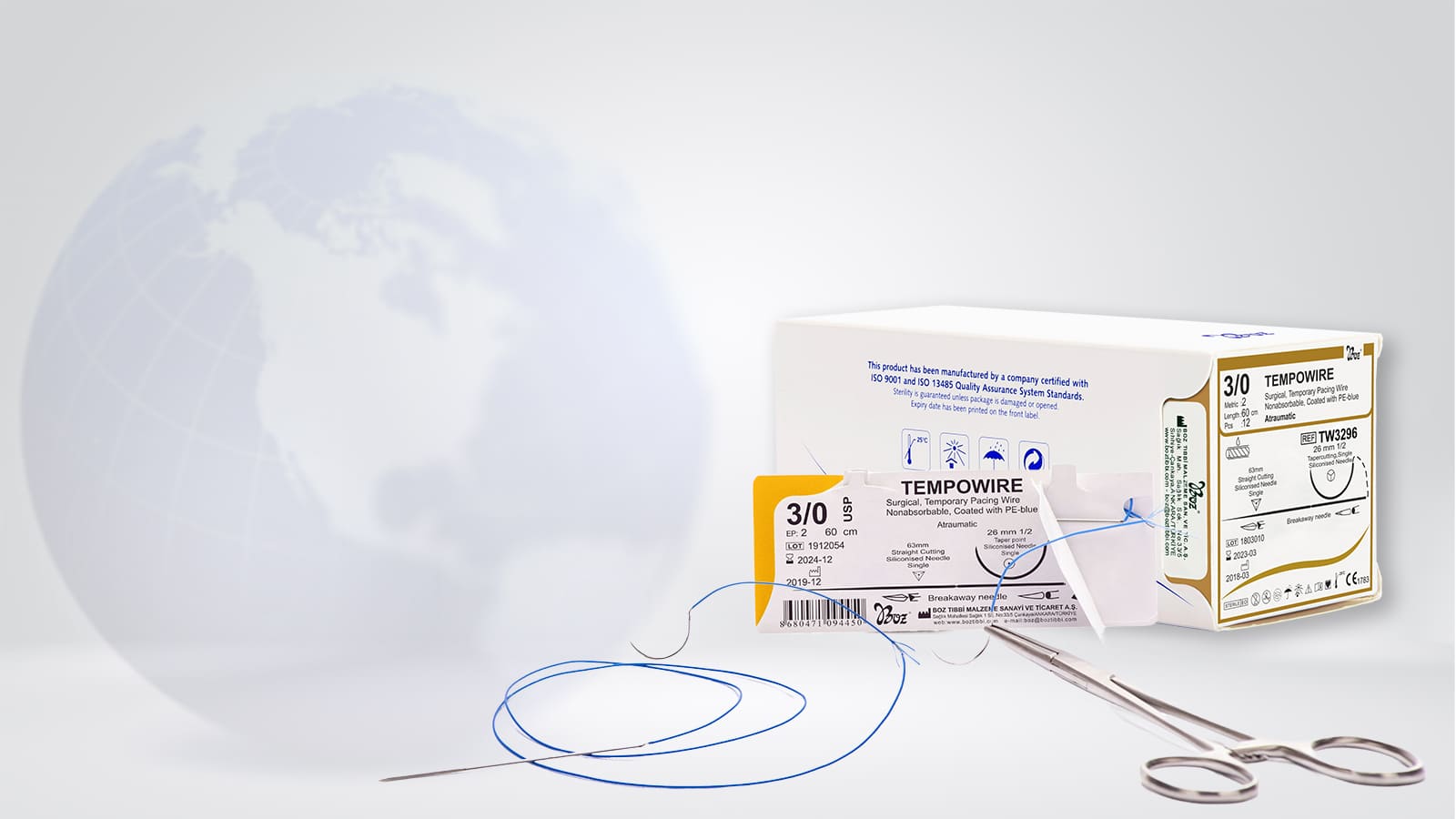
Please check the custom sterile package before opening. If the package is damaged, do not use the pace wire. Please check the instructions regarding opening the sterile package in Figure 2 and 3. The content of the sterile package is sterile (see also Figure 2, indication statement 1). Carefully remove the products in the sterile package. The insulation color on the surgical pacing wire body can be used for identification purpose. Please check Figure 4 for atrial usage (indication statement 1).


Insert the curved needle through the myocardium (see Figure 4) in a direction so that the entry point of the surgical temporary pacing wire into the heart can subsequently align with the exit in the chest. This method might facilitate retraction of the temporary pace wire. It is important to properly position the electrode inside or on the myocardium (see also Figure 5).
Atrial Usage: Position the electrode inside the myocardium. This is ensure by slightly pulling the curved needle while pulling the wire inside the tissue.
Cut the wire removed from the myocardium and curved needle until the rest of it stays in the myocardium (see also Figure 6). During the high displacement risk period, the cutting procedure might not be applied immediately because the wire is cut after a short period. Discard the myocardial curved needle.
Hold the chest needle distally from the separation mark (see Figure 7) and insert it through the chest wall in a direction that will allow the pacing wire in the heart to align with the chest exit. Keep some part of the conductive wire inside the check to prevent any displacement due to patient movement. Using a needle driver for this procedure is recommended.
Break the chest needle from separation mark (see also Figure 8). Discard the chest needle.
Connect the wire connector pins to the external pulse generator, patient cable, or surgical cable, following the polarity below. Connect the pin to the negative pole (-). The neutral wires must be connected to the positive pole (+) of the external pulse generator.
When the surgical wire is no longer needed, carefully pull to remove the wire.

Bibliography
Tempowire – Cerrahi, Geçici Pace Teli, Emilemeyen, Mavi-LD-PE kaplı – Kullanma Talimatı – Prospektüs (t.y.). Boz Tıbbi Malzeme. Erişim adresi: https://www.boztibbi.com/wp-content/uploads/2023/01/tempowire-ifu.pdf.
Knowledge Base

Properties and Usage Areas of Powder Hemostats