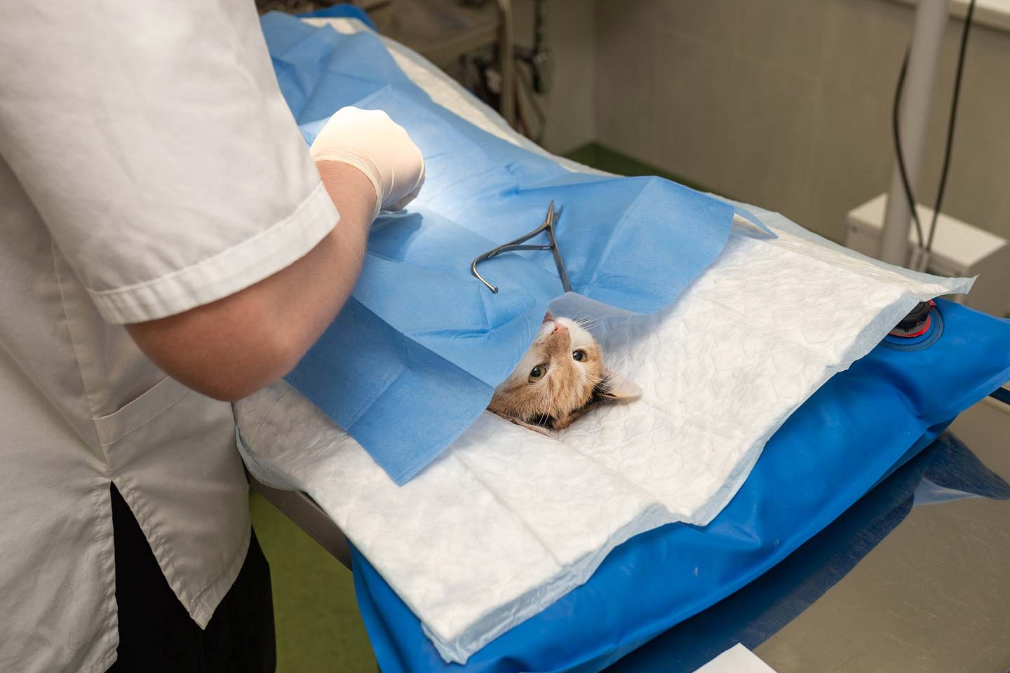
Sutures are used in the veterinary surgery to connect the damaged tissues or tissue incisions and for ligature applications. With the opportunities provided by the technology and science of today, there are numerous suture materials and each suture has unique usage areas and properties. The support provided by suture materials in scar and tissue recovery period plays an important role for the veterinary surgery. Please click here for the suture techniques used in the veterinary surgery. When choosing a suture, scar/incision conditions as well as physical, chemical and biologic properties of the sutures should be considered. In this article, we will talk about the properties of veterinary sutures.
What Are The Properties of Veterinary Surgery Suture Materials?
Surgical process that is implemented to connect the tissues and wounds in surgical operations or traumatic damages is called suturing (1). Sutures plays an important role in the veterinary surgery as they support tissue restoring and wound healing. Sutures in veterinary surgical operations are often preferred to close the tissues/wounds on skin, facia and muscle; digestive and urogenital system surgery, to stop the bleeding and in cardiovascular surgery (2).
Veterinary Surgery Suture Properties
Production Format
Most of the synthetic sutures are formed by polymerizing the liquid resin; silk suture formed from natural fibers is formed by bending and catguts are formed by stratification and bending after creating stripe forms (3).
Elasticity
The elasticity is the elongation of the suture by pulling and suture returning back to the previous position when set free. Elasticity is a desired property for the sutures. This elasticity also represents the strength. The power that the suture can withstand during flexing is defined as the strength. To prevent tissue tightening and cut due to oedema that might occur in the tissue after the suture is implanted to the wound might and tissue retract after the resorbation of oedema, the suture must get shorter to keep the tissues in the suitable shape and position (4,5).
Plasticity
This term represents the elongation of the suture materials with pulling and tugging but staying that way instead of returning to the initial position. In short, the suture elongates and cannot go back to the previous position. When this type of sutures are implanted to the tissue, the sutures will elongate without cutting or pressing the tissue which does not prevent the blood circulation around the wound. However, elongated suture due to tissue retraction after the resorbation of the oedama is unable to keep the tissues together. While most of the sutures have elastic property, some of them have plasticity (4,5).
Friction Surface
The suture surface must be uniform and smooth. However, sutures with too glossy and slippery surfaces are not preferred since these sutures do not hold the knots well (5). The granular suture surfaces are desired for knot security. The disadvantage of these sutures is that they cause trauma when passed through the tissue and cut the vein surface and cause thrombosis. These types of disadvantages are minimised by coating the sutures with silicon etc. materials. Multifilament (braided) sutures have more friction surface than monofilament (stripe/non-braided) sutures. Multifilament suture cause more trauma when passed through the tissues (6).
Memory
This represents that the suture cannot be changed easily. Sutures with high memory tend to go back to their packed format (4, 5) when the sutures are taken out of the package (5, 6) during and after manipulation (4,5).
Tensile Strength
The tensile strength of the suture represents the tensile breaking force (4, 5, 8). The suture tension starts to decrease after implantation. The tensile strength is related to the diameter of the suture and as the suture diameter increases, tensile strength increases. The weakest point of the suture is the knot. Therefore, tensile strength is measured from knotted suture (9). Knotted suture has the 3/2 strength of the unknotted suture (5). Each additional knot decreases the tensile strength of the suture by 30-40% and cause more foreign object to be on the tissue (6). The tensile strength especially plays an important role in tense areas such as linea alba (9). The results of Greenwald (10) that investigated the tensile strength of 10 different sutures with the same thickness for 6 weeks before and after in vivo incubation are given in Table 1.
| Suture Material | Tensile Strength Before Implantation (N/m2) | Tensile Strength 6 Weeks After Implantation (N/m2) | Tensile Strength 6 Weeks After Implantation Loss (%) |
|---|---|---|---|
| Silk | 0,159 | 0,125 | 22 |
| Polyglycolic Acid | 0,234 | Absorbed after 6 weeks | Almost non-existent |
| Polyester | 0,279 | 0,270 | Almost non-existent |
| Polyglycolic Lactic Acid | 0,329 | Absorbed after 6 weeks | Almost non-existent |
| Catgut | 0,351 | Completely absorbed after 6 weeks | Almost non-existent |
| Chromium Catgut | 0,393 | Completely absorbed after 6 weeks | Almost non-existent |
| Polypropylene | 0,577 | 0,479 | 17 |
| Polyglyconate | 0,612 | 0,316 | 49 |
| Polyamide 6 | 0,683 | 0,516 | 25 |
| Polyoxoanion | 0,784 | 0,332 | 58 |
Table 1 – Tensile Strength of Different Suture Materials for 6 Weeks Before and After In Vivo Incubation (10)
Usage Property
This represents a wide range of usage quality. This property is affected from suture handling, fraction coefficient, knot safety and memory (11).
Capillarity
This represents the liquid absorption by the surgical suture and keeping the absorbed liquid along the suture (4). Sutures with capillarity acts like a bougie (8, 12) and absorb the serum and bacteria in the implanted region and carry these along the suture (5, 8, 12). Most of the time the capillarity of the multifilament sutures are higher than monofilament sutures (4, 5, 12). Sutures with capillarity that are preferred on the skin permit microorganism transfer between external and internal environment and lead to contamination. The capillarity of the surgical sutures are minimised by coating with materials such as silicon, Teflon or resin (13).
Size
Surgical suture size are classified as “USP” in American Pharmacopeia and “EP” in European Pharmacopeia. The European codex uses metric system (5, 11, 12). Today, surgical suture sizes are often considered for USP classification. USP classification is done according to suture diameter, knot security and tension. Additionally, the classification changes for natural or synthetic structure and absorbable or non-absorbable nature of the suture. In EP classification, the millimetre width of the suture is considered as the measurement. The EP codes range between 0.1 and 20. Minimum radius can be found in terms of millimetre by dividing the code number with 10 (11).
| USP Size Codes | EP (Metric) Size Codes | Suture Material Diameter | |
|---|---|---|---|
| Natural Absorbable Suture Material | Natural and Synthetic Non-Absorbable/Synthetic Absorbable Suture Material | Non-Absorbable and Absorbable Suture Material | Min-Max (mm) |
| 8-0 | 8-0 | 0,4 | 0,04-0,049 |
| 7-0 | 7-0 | 0,5 | 0,05-0,069 |
| 6-0 | 6-0 | 0,7 | 0,07-0,099 |
| 6 | 6 | 9 | 0,90-0,99 |
| 5-0 | 5-0 | 1 | 0,10-0,14 |
| 5 | 5 | 8 | 0,80-0,89 |
| 4-0 | 4-0 | 1,5 | 0,15-0,19 |
| 4 | 4 | 7 | 0,70-0,79 |
| 3-0 | 3-0 | 2 | 0,2-0,24 |
| 3 | 3 | 6 | 0,60-0,69 |
| 2-0 | 2-0 | 2,5 | 0,25-0,29 |
| 2 | 2 | 5 | 0,50-0,59 |
| 1 | 1 | 4 | 0,40-0,49 |
| 11-0 | 0,1 | 0,01-0,019 | |
| 10-0 | 0,2 | 0,02-0,029 | |
| 9-0 | 0,3 | 0,03-0,039 | |
| 0 | 0 | 3 | 0,30-0,39 |
| 7 | 10 | 1,00-1,09 | |
Table 2 – Suture Size Classification (14)
Tissue Reaction
All sutures are a foreign substance for the tissues and lead to direct tissue reaction (8, 15). This reaction reaches the peak in 2-7 days depending on suture amount, type, configuration and implantation (4). Histologically, reaction implantation against the sutures are polymorphonuclear leucocyte between days 1-4, macrophage and fibroblast infiltration between days 4-7 and chronic inflammatory reaction and fibrose tissue formation after day 7 (15). In the meantime, fibrose capsule formation is observed around the sutures that are not absorbed on 28th day (6, 15) and inflammation continues until the sutures are completely absorbed for absorbable sutures (15).
Significant inflammatory reaction on the tissue decrease the strength of the wound against infection and delays wound healing (5). Sutures that lead to excessive tissue reaction might lead functional (capillary repair and ureteral anastomosis) or cosmetic problems (skin) due to excessive scar development (12). Tissue reaction is more in natural sutures compared to synthetic sutures and more in intestine and bladder than muscle and facia (6). Multifilament sutures cause more tissue reaction than monofilament sutures due to their capillary properties and increase the risk of infection (6, 15).
A study on rats showed that the most common inflammation reaction for wounds closed with chromium catguts, polyglycolic, silk and polypropylene were for catgut, silk, polypropylene and polyglycolic acid respectively (16). Kirpensteijn et al. worked on closing the dog skin incisions with polyglactin and poliglecaprone and found that poliglecaprone lead to less tissue reaction compared to polyglactin at the first stages of healing and there was no significant difference between two groups in the following stages of incision healing (17).
Physical Configuration
This defines whether the suture is monofilament or multifilament (4). Multifilament sutures are created by braiding or twisting multiple threads, monofilament sutures are formed by single thread (12). The threads in multifilament sutures prevent the bacteria to be phagocyted by the macrophage (6, 15). Multifilament sutures might absorb tissue fluid, swell and therefore lead the knots to be untied easily. Due to these properties, multifilament sutures lead to more tissue reactions (18).
Bibliography
(1) Samsar E, Akın F, Anteplioğlu H. (1996) Klinik Tanı Yöntemleri ve Genel Cerrahi. “6.Baskı” Tamer Matbaacılık. Ankara
(2) Tan R, Bell R, Dowling B, Dart A (2003) Suture Materials; Composition and Applications in Veterinary Wound Repair, Aust Vet J, 81(3):140-45
(3) Katz AR, Mukherjee DP, Kaganov AL, Gordon S (1985) A New Synthetic Monofilament YYÜ Vet Fak Derg, 2006, 17 (1-2):37-44
Absorbable Suture Made From Polytrimethlyene Carbonate. Surg Gynecol Obstet, 161(3): 213-22
(4) Moy RL, Lee A, Zalka A (1991): Commonly Used Suture Materials in Skin Surgery, Am Fam Physician, 44(6):2123-8
(5) Terhune, M (2002): Materials for wound closure, http://www.emedicine.com
(6) Monnet, E (2002): New Suture Materials Offer More Options for Wound Closures, The Newsmagazine of Veterinery Medicine, Oct, 1
(7) Leapar ,DJ (2001): Wound closure, EWMA, 1(2):19-24
(8) Niles J, Williams J (1999): Suture Materials and Patterns, In Practice, 21:308-20
(9) Taylor B, Bayat A (2003 ): Basic Plastic Surgery Techniques and Principles: Choosing the Right Suture Material, Student B.M.J., 11:140-41
(10) Greenwald D, Albear P, Gotlieb L (1994): Mechanical Comparison of 10 Suture Materials Before and After in Vivo İncubation, J Surg Res, 56: 372-77
(11) Henderson, RA (2005): The Veterinarian’s Suture Guide http://www.vetmed.auburn.edu
(12) Smeak, DD (1990): Selection and Use of Currently Available Suture Material, (in) Current Techniques in Small Animal Surgery, MJ Bojrab (Editor), chapter 3, 3nd Ed, Lea & Febiger , Philadelphia
(13) Katz AR, Mukherjee DP, Kaganov AL, Gordon S (1985): A New Synthetic Monofilament Absorbable Suture Made From Polytrimethlyene Carbonate Surg Gynecol Obstet, 161(3): 213-22
(14) Chu, CC (2001): Textile-Based Biomaterials for Surgical Aplications. (in) Polymeric Biomaterials Second Edition Revised and Expanded, S. Dumitriu, (Editör) Chapter 19, Marcel Dekker, New York.
(15) Sherbeeny, AM (2004): Needdles, Sutures and Knots Part III: Spesific Suture Materials, ASJOG, 1:167- 70
(16) Yaltirik M, Dedeoğlu K, Bilgic B, Koray M, Ersev H, Dulger O, Soley S (2003): Comparison of Four Different Suture in Soft Tissues of Rats, Oral Diseases, 9, 284-286.
(17) Kirpensteijn J, Maarschalkerweerd RJ, Koeman JP, Kooistra HS, Sluijs FJ (1997): Comparison of Two Suture Materials for Intradermal Skin Closure in Dog, Vet.Q.,19(1):20-22
(18) Parell, GJ 2003): Comparison of Absorbable with Nonabsorbable Sutures in Closure of Facial Skin Wounds, Arch Facial Plast Surg, 5, 6, 488-490